Ampulla of Vater
 From Wikipedia the free encyclopedia
| Ampulla of Vater | |
|---|---|
 A diagram of the biliary system. Note that the ampulla of Vater is behind the major duodenal papilla. | |
 The major duodenal papilla, seen on duodenoscopy at the time of ERCP. This is the protrusion of the ampulla of Vater into the duodenum. | |
| Details | |
| Identifiers | |
| Latin | ampulla hepatopancreatica, ampulla Vaterii |
| MeSH | D014670 |
| TA98 | A05.8.02.017 |
| TA2 | 3111 |
| FMA | 15076 |
| Anatomical terminology | |
The ampulla of Vater, hepatopancreatic ampulla or hepatopancreatic duct is the common duct that is usually formed by a union of the common bile duct and the pancreatic duct within the wall of the duodenum. This common duct usually features a dilation ("ampulla"). The common duct then opens medially into the descending part of the duodenum at the major duodenal papilla. The common duct usually measures 2-10mm in length.[1]
The ampulla of Vater is an important landmark halfway along the second part of the duodenum marking the transition from foregut to midgut.[citation needed]
Structure[edit]

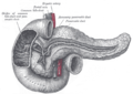
9. Gallbladder.
10–11. Right and left lobes of liver.
12. Spleen.
13. Esophagus.
14. Stomach.
15. Pancreas: 16. Accessory pancreatic duct, 17. Pancreatic duct.
18. Small intestine: 19. Duodenum, 20. Jejunum
21–22. Right and left kidneys.
The front border of the liver has been lifted up (brown arrow).[2]
Sphincters[edit]
Various smooth muscle sphincters regulate the flow of bile and pancreatic juice through the ampulla: the sphincter of the pancreatic duct, the sphincter of the bile duct, and the sphincter of Oddi.[3]
Variation[edit]
The common bile duct and pancreatic duct may sometimes unite outside the duodenal wall, creating an unusually long common duct. The two ducts may also drain into the duodenum separately, or may fuse yet retain their separate lumens separated by a septum.[1]
Clinical significance[edit]
Thomas' sign is the production of silver stools and can be indicative of cancer of the Ampulla of Vater. The silver-colored stool is a combination of the white stool of obstructive jaundice combined with black stool of melena or bleeding. It was first described in the British Medical Journal by Dr. H. Ogilvie in 1955.[4]
Etymology[edit]
The eponymic term "ampulla of Vater" is named after Abraham Vater (1684–1751),[5] a German anatomist who first published a description of it in 1723.[6]
Additional images[edit]
-
 The pancreatic duct.
 -
 Carcinoma of Ampulla


References[edit]
- ^ a b Standring, Susan (2020). Gray's Anatomy: The Anatomical Basis of Clinical Practice (42nd ed.). New York. p. 1219. ISBN 978-0-7020-7707-4. OCLC 1201341621.
{{cite book}}: CS1 maint: location missing publisher (link) - ^ Standring S, Borley NR, eds. (2008). Gray's anatomy : the anatomical basis of clinical practice. Brown JL, Moore LA (40th ed.). London: Churchill Livingstone. pp. 1163, 1177, 1185–6. ISBN 978-0-8089-2371-8.
- ^ Allescher, H. D. (1989). "Papilla of Vater: Structure and Function". Endoscopy. 21 (S 1): 324–329. doi:10.1055/s-2007-1012982. PMID 2691236. S2CID 38457896. Archived from the original on June 2, 2018. Retrieved March 27, 2020 – via www.thieme-connect.de.
- ^ Ogilvie, H (1955). "Thomas's sign, or the silver stool in cancer of the ampulla of Vater". Br Med J. 1 (4907): 208. doi:10.1136/bmj.1.4907.208. PMC 2060824. PMID 13219383.
- ^ Lerch, MM; Domschke, W (2000). "Abraham Vater of the ampulla (papilla) of Vater". Gastroenterology. 118 (2): 379. doi:10.1016/s0016-5085(00)70243-5. PMID 10691372.
- ^ Vater A. Dissertation in auguralis medica, poes diss. Qua Scirris viscerum dissert, C.S. Ezlerus. Vol 70. 1723.
- "Ampulla, hepatopancreatic." Stedman's Medical Dictionary, 27th ed. (2000). ISBN 0-683-40007-X
- Moore, Keith L. and Arthur F. Dalley. Clinically Oriented Anatomy, 4th ed. (1999). ISBN 0-683-06141-0