Thoracic splanchnic nerves
 From Wikipedia the free encyclopedia
| Thoracic splanchnic nerves | |
|---|---|
 The right sympathetic chain and its connections with the thoracic, abdominal, and pelvic plexuses. (Greater and lesser splanchnic nerves labeled at left.) | |
 Abdominal portion of the sympathetic trunk, with the celiac and hypogastric plexuses. (Greater splanchnic and lowest splanchnic labeled at upper left. Greater splanchnic and lesser splanchnic labeled at upper right.) | |
| Details | |
| From | thoracic ganglia |
| Innervates | greater splanchnic nerve: celiac ganglia lesser splanchnic nerve: superior mesenteric ganglion and aorticorenal ganglion least splanchnic nerve: renal plexus |
| Identifiers | |
| TA98 | A14.3.01.028 A14.3.01.032 A14.3.01.030 |
| TA2 | 6631, 6632, 6634 |
| FMA | 6280 |
| Anatomical terms of neuroanatomy | |
Thoracic splanchnic nerves are splanchnic nerves that arise from the sympathetic trunk in the thorax and travel inferiorly to provide sympathetic supply to the abdomen. The nerves contain preganglionic sympathetic fibers and general visceral afferent fibers.
Nerves[edit]
There are three main thoracic splanchnic nerves.[1]
| Name | Spinal Nerve Roots | Ganglia | Structure | Function |
|---|---|---|---|---|
| Greater splanchnic nerve | T5–T9 | T5–T9 T5–T10 | The greater splanchnic nerve travels through the diaphragm and enters the abdominal cavity. Its fibers synapse at the celiac ganglia.[4] The nerve contributes to the celiac plexus, a network of nerves located in the vicinity of where the celiac trunk branches from the abdominal aorta. | The greater splanchnic nerve modulates the activity of the enteric nervous system of the foregut. It stimulates contraction of the splanchnic vasculature, increasing blood pressure.[5] It also provides sympathetic innervation to the adrenal medulla, stimulating catecholamine release. It may provide sensory innervation to the pancreas.[1] |
| Lesser splanchnic nerve | T9–T12 | T9–T12 T9–T10 T10–T12 T10–T11 | The lesser splanchnic nerve travels inferiorly, lateral to the greater splanchnic nerve. Its fibers synapse with their postganglionic counterparts in the superior mesenteric ganglion, or in the aorticorenal ganglion.[4] | The lesser splanchnic nerve modulates the activity of the enteric nervous system of the midgut. |
| Least splanchnic nerve | T12 | T12–L2 T11–T12[6] | The least splanchnic nerve travels into the abdomen medial to the sympathetic trunk.[6] Its fibers synapse in the renal plexus.[4][6] |
Additional images[edit]
-
 Greater splanchnic nerve, seen in thoracic cavity seen from left side.
 -
 The celiac ganglia with the sympathetic plexuses of the abdominal viscera radiating from the ganglia.
 -
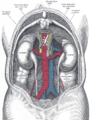 The relations of the viscera and large vessels of the abdomen. Seen from behind, the last thoracic vertebra being well raised.
 -
 Thoracic splanchnic nerves


References[edit]
- ^ a b c d e f g h Kline, Matthew T. (2007-01-01), Waldman, Steven D.; Bloch, Joseph I. (eds.), "chapter 169 – Radiofrequency Techniques", Pain Management, Philadelphia: W.B. Saunders, pp. 1411–1459, doi:10.1016/b978-0-7216-0334-6.50173-4, ISBN 978-0-7216-0334-6, retrieved 2020-11-23
- ^ a b c Moore, Keith (2018). Clinically Oriented Anatomy. Wolters Kluwer. pp. 59–61. ISBN 9781496347213.
- ^ a b thoraxlesson5 at The Anatomy Lesson by Wesley Norman (Georgetown University)
- ^ a b c Waxenbaum, Joshua A. (29 July 2021). "Anatomy, Autonomic Nervous System". StatPearls Publishing. PMID 30969667 – via Europe PMC.
- ^ Bapna, Anisha; Adin, Christopher; Engelman, Zoar J.; Fudim, Marat (2020-08-01). "Increasing Blood Pressure by Greater Splanchnic Nerve Stimulation: a Feasibility Study". Journal of Cardiovascular Translational Research. 13 (4): 509–518. doi:10.1007/s12265-019-09929-7. ISSN 1937-5395. PMID 31691154. S2CID 207896247.
- ^ a b c Gray's anatomy : the anatomical basis of clinical practice. Susan Standring (41st ed.). Philadelphia: Elsevier. 2015. ISBN 978-0-7020-5230-9. OCLC 920806541.
{{cite book}}: CS1 maint: others (link)
External links[edit]
- Anatomy figure: 21:04-07 at Human Anatomy Online, SUNY Downstate Medical Center – "The position of the right and left vagus nerves, and sympathetic trunks in the mediastinum."
- Anatomy photo:40:10-0102 at the SUNY Downstate Medical Center – "Posterior Abdominal Wall: The Celiac Plexus"
- figures/chapter_30/30-4.HTM: Basic Human Anatomy at Dartmouth Medical School
- figures/chapter_32/32-6.HTM: Basic Human Anatomy at Dartmouth Medical School
Anatomy of the autonomic nervous system | |||||
|---|---|---|---|---|---|
| Head |
| ||||
| Neck |
| ||||
| Thorax |
| ||||
| Abdomen |
| ||||
| Pelvis |
| ||||