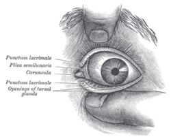
Lacrimal caruncle
 From Wikipedia the free encyclopedia
| Lacrimal caruncle | |
|---|---|
 Front of left eye with eyelids separated. Caruncula visible and labeled at left. | |
| Details | |
| System | Eye |
| Identifiers | |
| Latin | caruncula lacrimalis |
| TA98 | A15.2.07.049 |
| TA2 | 6844 |
| FMA | 77672 |
| Anatomical terminology | |
The lacrimal caruncle, or caruncula lacrimalis, is the small, pink, globular nodule at the inner corner (the medial canthus) of the eye.[1] It consists of tissue types of neighbouring eye structures. It may suffer from lesions and allergic inflammation.
Structure
[edit]The lacrimal caruncle is found at the medial canthus of the eye.[1] It consists of skin, hair follicles, sebaceous glands, sweat glands, accessory lacrimal tissue and other tissues that are present in the skin and accessory lacrimal glands.[1][2] Its non-keratinized epithelium resembles the conjunctival epithelium.[2]
Clinical significance
[edit]Lesions
[edit]The lacrimal caruncle may have a lesion.[3] This can have any one of a number of causes, which may be difficult to diagnose.[3] Cancer is a rare cause.[3][4] These lesions include papillomas and oncocytomas.[4]
Allergies
[edit]With ocular allergies, the lacrimal caruncle and the plica semilunaris of the conjunctiva may be inflamed and pruritic (itchy) due to histamine release in the tissue and tear film.
Other diagnoses
[edit]Sweat glands and oil glands are contained in the caruncle of the eye (lacrimal caruncle in medial canthus). As with all oil glands, lacrimal caruncles can become clogged, causing a pimple, whitehead, or pustule beneath the skin. Clogged oil and sweat glands in the caruncle can affect tear ducts. Treatment for dry eyes due to clogged glands includes refraining from rubbing the eyes and rinsing the eyes with clear water frequently during the day, either with clean hands or a spray faucet. Additionally, one can use a warm damp cloth on the eye, which will help the clogged pore to open up and release some pressure. Anti-bacterial eye drops may also be prescribed. If the pustules enlarge, an oral antibiotic may be prescribed. If lesions such as cysts form, they must be surgically drained; this operation is rarely necessary. [5] If it affects the tear sac it may be dacryocystitis.
References
[edit]- ^ a b c Okumura, Yuta; Takai, Yoshiko; Yasuda, Shunsuke; Terasaki, Hiroko (2017). "Bilateral lacrimal caruncle lesions". Nagoya Journal of Medical Science. 79 (1): 85–90. doi:10.18999/nagjms.79.1.85. ISSN 0027-7622. PMC 5346624. PMID 28303065.
- ^ a b Miura-Karasawa, Maria; Toshida, Hiroshi; Ohta, Toshihiko; Murakami, Akira (27 April 2018). "Papilloma and sebaceous gland hyperplasia of the lacrimal caruncle: a case report". International Medical Case Reports Journal. 11: 91–95. doi:10.2147/IMCRJ.S162528. PMC 5927183. PMID 29731668.
- ^ a b c Santos, Arturo; Gómez-Leal, Alfredo (1994-05-01). "Lesions of the Lacrimal Caruncle: Clinicopathologic Features". Ophthalmology. 101 (5): 943–949. doi:10.1016/S0161-6420(94)31233-4. ISSN 0161-6420. PMID 8190485.
- ^ a b Kapil, Jyoti P.; Proia, Alan D.; Puri, Puja Kumari (May 2011). "Lesions of the Lacrimal Caruncle With an Emphasis on Oncocytoma". The American Journal of Dermatopathology. 33 (3): 227–235. doi:10.1097/DAD.0b013e3181d9b56d. ISSN 0193-1091. PMID 21522047. S2CID 22955802.
- ^ Levy, J., Ilsar, M., Deckel, Y. et al. Lesions of the caruncle: a description of 42 cases and a review of the literature. Eye 23, 1004–1018 (2009). https://doi.org/10.1038/eye.2008.316
Additional images
[edit]- Extrinsic eye muscle. Nerves of orbita. Deep dissection
- Small white lesion on lacrimal caruncle, most likely a cyst

