Oncocytoma
 From Wikipedia the free encyclopedia
| Oncocytoma | |
|---|---|
| Specialty | Oncology |
| Symptoms | 4 |
An oncocytoma is a tumor made up of oncocytes, epithelial cells characterized by an excessive amount of mitochondria, resulting in an abundant acidophilic, granular cytoplasm.[1][2] The cells and the tumor that they compose are often benign but sometimes may be premalignant or malignant.
Presentation[edit]
An oncocytoma is an epithelial tumor composed of oncocytes, large eosinophilic cells having small, round, benign-appearing nuclei with large nucleoli.[citation needed]
Oncocytoma can arise in a number of organs.[citation needed]
Renal oncocytoma[edit]
Renal oncocytoma is thought to arise from the intercalated cells of collecting ducts of the kidney. It represents 5% to 15% of surgically resected renal neoplasms.[citation needed]
Salivary gland oncocytoma[edit]

An salivary gland oncocytoma (also known as an oxyphilic adenoma) is a well-circumscribed, benign neoplastic growth comprising about one percent of all salivary gland tumors. The histopathology is marked by sheets of large, swollen polyhedral epithelial oncocytes, which are granular acidophilic parotid cells with centrally located nuclei. The granules are created by the mitochondria.[citation needed]
Symptoms[edit]
Salivary gland oncocytomas, 85 to 90 percent of which are located in the parotid gland, are firm, slowly growing, painless masses of less than 4 cm and may be bilateral. They are most common in females age 70 to 80.[citation needed]
Thyroid oncocytoma[edit]
Thyroid oncocytomas (also known as Hürthle cell tumours) can be benign (adenomas) or malignant (carcinomas). Grossly, oncocytic adenomas are encapsulated, solid nodules with a characteristic brown cut surface. The gross appearance of a minimally invasive oncocytic carcinoma is indistinguishable to that of an adenoma, while widely invasive oncocytic carcinomas are obviously invasive macroscopically and display pervasive vascular invasion with multifocal involvement of the thyroid gland. There are no reliable cytologic features which distinguish oncocytic adenomas from carcinomas and the only criterion for a diagnosis of malignancy is the identification of transcapsular or vascular invasion.[citation needed]
Symptoms[edit]
Patients with thyroid oncocytomas present with a thyroid nodule, usually with normal thyroid function. If the tumor is big or invasive, there may be other symptoms such as difficulty swallowing or talking.[citation needed]
Additional images[edit]
-

-
 Micrograph of a renal oncocytoma. H&E stain.
 -
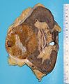 Gross appearance of the cut surface of a nephrectomy specimen containing a renal oncocytoma. Note the rounded contour, the mahogany colour and the central scar.


See also[edit]
References[edit]
- ^ Coburn V, Radfar A, Snook D, Mahalingam M (April 2007). "Cutaneous oncocytoma - a report of three cases and review of the literature". Journal of Cutaneous Pathology. 34 (4): 355–359. doi:10.1111/j.1600-0560.2006.00620.x. PMID 17381809. S2CID 19955625.
- ^ "Atlas of Genetics and Cytogenetics in Oncology and Haematology - Thyroid:oncocytic tumors". Retrieved 2009-02-01.